


With severe internal diseases, poor nutrition, as well as with age, the growth of the nail slows down, its structure undergoes changes. Only a doctor can accurately determine the cause of the violation based on the results of microscopic tests and examinations.
But to get an idea of what happens to the nails of the legs or hands, you can use the photo with fungal diseases of different types.
Causes of nail deformation
Molds, yeast-like fungi and dermatophyte fungi cause infectious nail diseases (onychomycosis), which show similar symptoms.
All types of nail fungus or nail deform the nail plate, change its transparency, brightness, color, this variety can be seen in the photos presented.
Nail changes occur not only with onychomycosis, but also with lesions, chronic paronychia (inflammation of the nail fold), psoriasis, eczema of the hands, dermatitis. Before concluding that there is a fungal infection, it is necessary to consider all possible options.
Signs of yeast infection
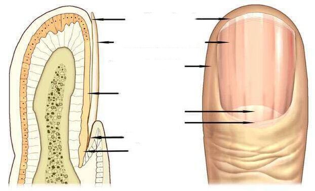
The most informative signs of fungal infection are changes in the color of the nail plate, presence of nail detachment, superficial changes - transversal, longitudinal grooves in the nail plate, point depressions, thickening, nail destruction.
The pink color of a healthy nail is determined by the transparency of the nail plate and the blood vessels visible through it. In onychomycosis, the nail loses its transparency, the color becomes brownish, yellow, less often green, black.
Candida fungi and dermatophytes cause onycholysis - the separation of the affected part of the nail. When infected with dermatophytes, onycholysis is observed at the remote edge of the nail and, when infected with Candida, the nail is behind the nail bed at the base, in the area of the crescent moon.
A symptom of a candidal fungus may be inflammation of the lateral periungual ridges - paronychia. This disease has bacterial forms caused by streptococci and staphylococci, as well as non-infectious forms - eczema, psoriasis, systemic vasculitis.
When the toenails are affected by the fungus Trichophyton rubrum, the plaque is affected, as you can see in the photo, the roller is not affected by the infection. The plaque becomes yellowish, thickens strongly, the accumulated fungal masses are well distinguished under it.
Nail fungus due to dermatophyte infection
In 95% of all nail fungus cases, the disease is caused by dermatophytes Trichophyton rubrum and Trichophyton mentagrophytes.
Trichophyton rubrum infection
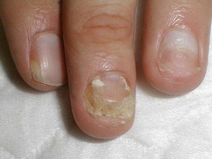
Onychomycosis begins when the fungus penetrates under the nail plate from the free edge. The fungal infection is indicated by the appearance of a yellowish spot, an irregular and crumbling surface of the distal (remote) edge of the nail in the spot of the spot.
distal-lateral formof Trichophyton rubrum dermatophyte fungus infection is common. In the photo, it is possible to observe that the spot caused by the introduction of the fungus is located along the lateral nailfold nail fold.
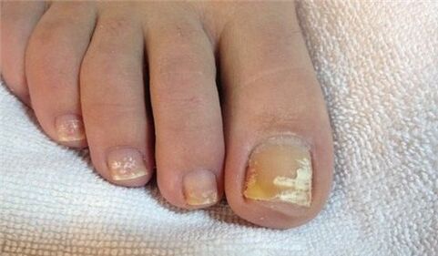
The fungus Trichophyton rubrum usually affects the big toes, causing hyperkeratosis - an accumulation of fungi between the nail plate and the nail bed, which looks like a loose yellowish mass in the photo.
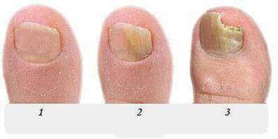
At this stage, the fungus occupies an insignificant part of the nail, as in the photo presented, and with the help of local treatment it is possible to face incipient onychomycosis.
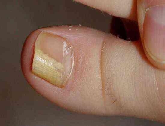
Without treatment, the stain grows, gradually affects the entire edge of the nail and then moves to the half moon. In the photo, the nail fungus looks like yellowish stripes directed to the growth zone of the nail plate.
With thedistal shape of the nail fungus, which is usually found on the big toes, a yellowish spot of infection appears on the distal edge of the nail, in its central part, as seen in thePhotograph.
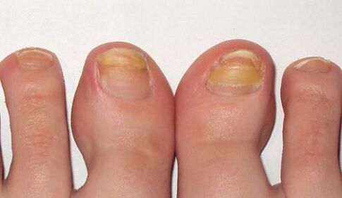
In the advanced stage of the fungus on the legs, several nails are affected, as in the photo, and treatment is no longer limited to local remedies and pills. In addition to antifungal agents, the nail is subjected to cleaning of the hardware, for the total or partial removal of the nail plate.
Long-term therapy with the use of all known antifungals and treatments should be performed on the leg, caused by Trichophyton rubrum, with hyperkeratosis, as seen in the photo.
The fungal infection with total nail damage spreads throughout the nail plate area, the nail is completely destroyed.
Infection by another representative of dermatophytes, the fungus Trichophyton mentagrophytes, can also lead to a total fungal infection of the nail.
Trichophyton mentagrophytes infection
With the total defeat of the nail with the fungus Trichophyton mentagrophytes, the nail plate is deformed, the photo shows that it thickens, changes its structure, falls, yellow spots appear on its entire surface.
Infection of the nail by this dermatophyte usually causes superficial white onychomycosis of the big toe, less frequently of the big toe.
This fungus practically does not occur on the nails of the hands, usually causes interdigital dermatophytosis on the legs, as in the photo, and requires simultaneous treatment of the skin of the feet and nails.
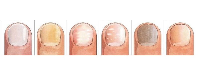
A symptom of fungal infection on the nails, usually on the feet, are white patches of various sizes, as in the photo, which resemble leukonychia - a disease of the nail plate itself.
But unlike leukonychia, where the white spots are caused by the appearance of air bubbles in the nail layers, the white spots in a fungal infection are the result of the activity of Trichophyton mentagrophytes.
Rarely, superficial white onychomycosis is caused by fungi; in AIDS, the causative agent of this type of fungus may be Trichophyton rubrum and affect the nails of the feet and hands.
Nail changes due to Candida infection
The fungus usually occurs in women, affects the nails of the active hand, which is most often in contact with water.
For Candida onychomycosis, the proximal form of the infection is characteristic, in which the fungus first affects the nail fold at the base of the nail, then penetrates the growth zone and the nail bed. Then, it moves gradually along the nail, from the base to the edge, capturing an increasingly larger area of the nail plate.
The causative agent of Candida onychomycosis is Candida albicans. This fungus invades the nails of the feet and hands, spreading from the area of the half moon at the base of the nail plate, to the free edge, as can be seen in the photo.
A sign of Candida nail infectionalbicans is inflammation of the nail fold (paronychia), separation of the cuticle from the nail plate, pain, pus secretion when a bacterial infection occurs.
Candida albicans is able to penetrate the nail and its free edge. In this case, they speak of the distal form of the infection, which is usually associated with cutaneous candidiasis.
The treatment of candida fungus on the nails of the hands and feet with damage in more than half of the nail plate area, as in the photo, includes not only the fight against onychomycosis, but also measures to reduce candida activityin the natural reservoirs of its storage - the intestines, the oral cavity, the genital mucosa. . .
Fungal infestation
Molds cause fungi much less frequently than Candida or dermatophytes. The main symptom of nail infection with mold is, as you can see in the photo, inchange the color of the nail plate to blue, black, greenish.
The signs of mold on the nails can be dark spots, spots on the nail plate or, as in the photo, a longitudinal black band.
Preparations against fungi
Antifungal agents with fluconazole, ketoconazole, terbinafine, itraconazole, griseofulvin are used to treat nail fungus caused by dermatophytes, as in this photo.
Antifungal agents with terbinafine are effective for dermatophyte infections.
Antifungal agents with voriconazole are highly active against dermatophytes.
It isused andto treat nail moldon the feet, hands andagainst candida yeast. The spectrum of action includes fungi like Aspergillum, Fusarium, Penicillium.
Itraconazole-based preparations deal with mold.
Fungal nail diseases
A grayish tonesometimes appears on thenail with eczema. In this case, the nail plate may move away from the nail bed, which is observed with a fungus.
Externally very similar to onychomycosismanifestations of psoriasis. With this disease, not onlychanges color, but alsothe nail plate thickens.
Point depressions are found on its surface, the separation of the nail plate from the nail bed is observed. But there are differences in relation to the fungus: in psoriasis, the highlighted and healthy parts of the toenail are separated by a yellowish pink band over time.
The bluish coloraffects thenail with pseudomonas nail infection. Frequent mechanical friction of the nail fold causes the appearance of superficial streaks, waviness of the nail.
White spots of leukonychia, whose appearance isassociated with metabolic disorders, can also be confused with a superficial white fungus with a large area of the spot.
Changes in the color and shape of the nail causing injuries. Thumbs are at greater risk. The nail with a lesion, as well as with a fungus, thickens and darkens.

The difference between the lesion and the fungus is that changes during the lesion are noticed only in the injured finger, the nails of the other fingers remain unchanged, they do not infect the sick finger, as in onychomycosis.
The consequence of trauma can be the partial separation of the nail from the nail bed, the formation of a cavity that, under unfavorable conditions, is quickly colonized by fungi.
The nail plate can be separated from the nail bed under the influence of light (photoonicolysis), with iron deficiency anemia, hormonal diseases. Division, nail loss occurs with lichen erythematosus, bullous dermatoses, nail trauma.
But you can finally be sure that the conclusion is correct and start the treatment, you will only be able to consult a dermatologist - specialist in skin diseases, or a mycologist - doctor who treats fungal diseases.